Pfam Domains mapped on to the structure: 1G85
No.
Chain ID
Pfam ID
Pfam Description
Linkout - Pfam
Linkout - CDD
1
A
PF00061
Lipocalin / cytosolic fatty-acid binding protein family
PF00061
PF00061
Conserved Domain Database Superfamily Annotations: 1G85
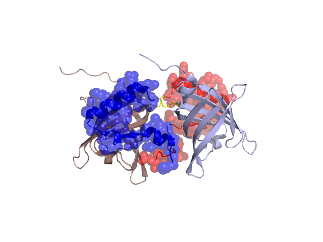
Structural Details of PDB entry 1G85
Structural Details of PDB entry 1G85
PDBid Chains Hinge Swapped Domain
1G85
A,B
A:121-123,B:121-123
A:124-159,B:124-157
Swapped-domain interface residues and interactions:
Chains Residues
A
19 , 20 , 21 , 22 , 23 , 24 , 25 , 27 , 100 , 117 , 118 , 124 , 125 , 128 , 129 , 131 , 132 , 133 , 135 , 139 , 141 , 145 , 146 , 147 , 148 , 149 , 150 , 152 , 154 , 155 , 156 , 157 , 158 , 159 ,
B
19 , 20 , 21 , 22 , 23 , 24 , 25 , 27 , 37 , 64 , 100 , 117 , 118 , 120 , 124 , 125 , 128 , 129 , 131 , 132 , 133 , 135 , 139 , 141 , 145 , 146 , 147 , 148 , 149 , 150 , 152 , 153 , 156 ,
Non-swapped-domain interface residues and interactions:
Chains Residues
A
18 , 30 , 37 , 39 , 92 , 93 , 97 , 98 , 102 , 114 , 116 , 120 , 122 , 123 ,
B
18 , 30 , 32 , 39 , 57 , 59 , 92 , 93 , 97 , 98 , 102 , 114 , 116 , 122 , 123 ,
Mutations in critical regions:
Chains
Hinge
Domain swapped interface Non-swapped interface Swapped Domain
A No mutation GLY(117)GLU, ASN(154)ASP, No mutation ASN(154)ASP, B No mutation GLY(117)GLU, No mutation ASN(154)ASP,
HIDE output:
JMOL Visualization:
2D-plot:
JOY Structural annotation for hinge hinge and swapped domain:
JOY output: