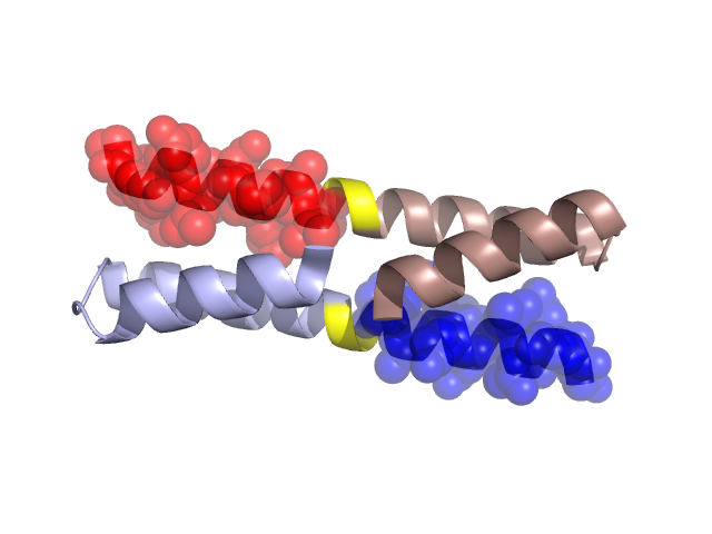
Structural Details of PDB entry 1G6U Structural Details of PDB entry 1G6U PDBid Chains Hinge Swapped Domain 1G6U A,B A:33-34,B:33-34 A:35-48,B:35-48 Swapped-domain interface residues and interactions: Chains Residues A 2, 5, 25, 35, 36, 39, 40, 42, 43, 46, 47, 48, B 25, 35, 36, 39, 40, 42, 43, 46, 47, Non-swapped-domain interface residues and interactions: Chains Residues A 1, 8, 12, 17, 21, 28, 32, B 1, 2, 5, 8, 12, 17, 18, 21, 28, 32, Swapped domains are represented using trasperent spheres. Non-swapped part is represented using light color and cartoon representation. Hinge region is shown in yellow color. Mutations in critical regions: Chains Hinge Domain swapped interface Non-swapped interface Swapped Domain ANo mutationNo mutationNo mutationNo mutation BNo mutationNo mutationNo mutationNo mutation HIDE output: Homologues found through HIDE algorithm JMOL Visualization: 2D-plot: A:1G6U B:1G6U JOY Structural annotation for hinge hinge and swapped domain: Hinge Swapped domain JOY output: ali file:1G6U.ali atm file:1G6U.atm cof file:1G6U.cof hbd file:1G6U.hbd html file:1G6U.html pdb file:1G6U.pdb ps file:1G6U.ps psa file:1G6U.psa rtf file:1G6U.rtf sst file:1G6U.sst tem file:1G6U.tem