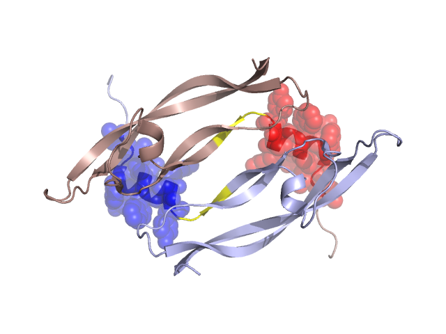
Structural Details of PDB entry 1FZV
Structural Details of PDB entry 1FZV
PDBid Chains Hinge Swapped Domain
1FZV
A,B
A:35-38,B:35-38
A:23-34,B:23-34
Swapped-domain interface residues and interactions:
Chains Residues
A
23 , 24 , 25 , 26 , 29 , 30 , 32 , 33 , 41 , 58 , 60 , 61 , 62 , 86 , 87 , 88 ,
B
23 , 24 , 25 , 26 , 29 , 30 , 32 , 33 , 41 , 58 , 60 , 61 , 62 , 86 , 87 , 88 ,
Non-swapped-domain interface residues and interactions:
Chains Residues
A
21 , 22 , 36 , 38 , 39 , 40 , 57 , 59 , 67 , 68 , 69 , 85 , 90 , 100 , 102 , 117 ,
B
21 , 22 , 36 , 38 , 39 , 40 , 57 , 59 , 67 , 68 , 69 , 85 , 90 , 100 , 102 ,