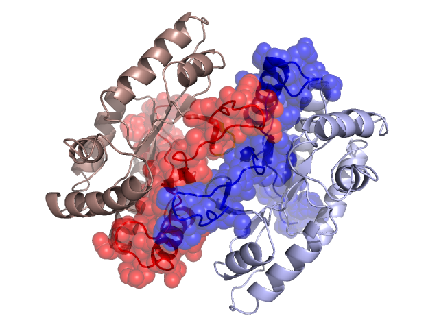
Pfam Domains mapped on to the structure: 1FVP No. Chain ID Pfam ID Pfam Description Linkout - Pfam Linkout - CDD 1 A PF00296 Luciferase-like monooxygenase PF00296 PF00296 Conserved Domain Database Superfamily Annotations: 1FVP No. PDB ID PSSM ID CDD Accession Superfamily Short Name Linkout - CDD 1 1FVP 212284 Flavin_utilizing_monoxygenases superfamily N - Structural Details of PDB entry 1FVP Structural Details of PDB entry 1FVP PDBid Chains Hinge Swapped Domain 1FVP A,B A:69-71,B:69-71 A:1-68,B:1-68 Swapped-domain interface residues and interactions: Chains Residues A 12, 13, 14, 22, 25, 26, 29, 32, 35, 36, 43, 44, 45, 47, 49, 51, 52, 54, 56, 57, 60, 61, 62, 63, 65, 67, 69, 70, 155, B 12, 14, 22, 25, 26, 29, 32, 35, 36, 43, 44, 45, 46, 47, 51, 52, 54, 55, 56, 57, 60, 61, 62, 63, 65, 67, 69, 70, Non-swapped-domain interface residues and interactions: Chains Residues A 72, 88, 90, 231, B 71, 72, 88, 90, 155, Swapped domains are represented using trasperent spheres. Non-swapped part is represented using light color and cartoon representation. Hinge region is shown in yellow color. Mutations in critical regions: Chains Hinge Domain swapped interface Non-swapped interface Swapped Domain ANo mutationNo mutationNo mutationPRO(64)SER, BNo mutationNo mutationNo mutationPRO(64)SER, HIDE output: Homologues found through HIDE algorithm JMOL Visualization: 2D-plot: A:1FVP B:1FVP JOY Structural annotation for hinge hinge and swapped domain: Hinge Swapped domain JOY output: ali file:1FVP.ali atm file:1FVP.atm cof file:1FVP.cof hbd file:1FVP.hbd html file:1FVP.html pdb file:1FVP.pdb ps file:1FVP.ps psa file:1FVP.psa rtf file:1FVP.rtf sst file:1FVP.sst tem file:1FVP.tem