Pfam Domains mapped on to the structure: 1FJH
No.
Chain ID
Pfam ID
Pfam Description
Linkout - Pfam
Linkout - CDD
1
A
PF13561
Enoyl-(Acyl carrier protein) reductase
PF13561
PF13561
Conserved Domain Database Superfamily Annotations: 1FJH
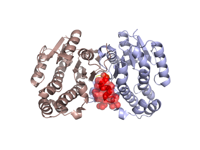
Structural Details of PDB entry 1FJH
Structural Details of PDB entry 1FJH
PDBid Chains Hinge Swapped Domain
1FJH
A,B
A:242-246,B:242-246;A:1242-1246,B:1242-1246
A:247-257,B:247-257
Swapped-domain interface residues and interactions:
Chains Residues
A
247 , 249 , 250 , 251 , 253 , 254 , 255 , 257 ,
B
1122 , 1164 , 1167 , 1168 , 1170 , 1171 , 1238 , 1239 , 1240 ,
Non-swapped-domain interface residues and interactions:
Chains Residues
A
118 , 122 , 163 , 164 , 167 , 168 , 170 , 171 , 174 , 177 , 179 , 187 , 209 , 211 , 212 , 213 , 215 , 217 , 221 , 224 , 225 , 228 , 233 , 235 , 236 , 237 , 238 , 239 , 240 , 241 , 242 , 243 , 244 , 245 , 246 ,
B
1106 , 1117 , 1118 , 1163 , 1174 , 1177 , 1179 , 1212 , 1213 , 1215 , 1217 , 1221 , 1224 , 1225 , 1228 , 1233 , 1235 , 1236 , 1237 , 1241 , 1242 , 1243 , 1244 , 1245 , 1246 , 1247 , 1249 , 1250 , 1251 , 1253 , 1254 , 1255 ,
Mutations in critical regions:
Chains
Hinge
Domain swapped interface Non-swapped interface Swapped Domain
A No mutation No mutation No mutation No mutation B No mutation No mutation No mutation No mutation B No mutation No mutation No mutation No mutation
HIDE output:
JMOL Visualization:
2D-plot:
JOY Structural annotation for hinge hinge and swapped domain:
JOY output: