Home BrowseSearch Help Group

Pfam Domains mapped on to the structure: 1FIP

No. Chain ID Pfam ID Pfam Description Linkout - Pfam Linkout - CDD
1 A PF02954 Bacterial regulatory protein, Fis family PF02954 PF02954
Structural Details of PDB entry 1FIP

Structural Details of PDB entry 1FIP

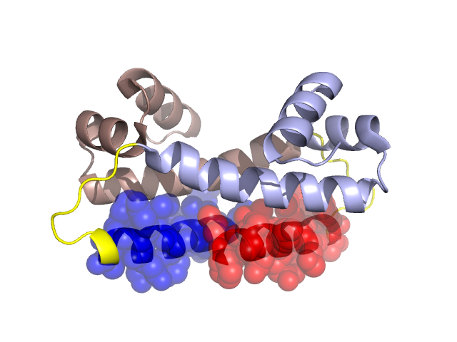
PDBid Chains Hinge Swapped Domain
1FIP A,B A:41-48,B:41-48 A:26-40,B:26-40

Swapped-domain interface residues and interactions:

Chains Residues
A 27, 28, 30, 31, 32, 34, 35, 36, 39, 57, 58, 61, 62, 65,
B 27, 28, 30, 31, 32, 34, 35, 39, 57, 58, 61, 62, 65,

Non-swapped-domain interface residues and interactions:

Chains Residues
A 42, 47, 48, 50, 51, 54, 55, 59, 60, 64, 66, 69, 76, 79, 80, 81, 82, 83, 91, 98,
B 42, 47, 48, 50, 51, 54, 55, 59, 60, 64, 66, 69, 79, 80, 81, 82, 83, 91,

Swapped domains are represented using trasperent spheres.
Non-swapped part is represented using light color and cartoon representation.
Hinge region is shown in yellow color.

Mutations in critical regions:

Chains Hinge Domain swapped interface Non-swapped interface Swapped Domain
ANo mutationALA(61)PRO, No mutationNo mutation
BNo mutationALA(61)PRO, No mutationNo mutation

HIDE output:

Homologues found through HIDE algorithm

JMOL Visualization:

Jmol_logo            Jmol_logo            Jmol_logo

2D-plot:

A:1FIP B:1FIP

JOY Structural annotation for hinge hinge and swapped domain:

Hinge Swapped domain

JOY output:

ali file:1FIP.ali
atm file:1FIP.atm
cof file:1FIP.cof
hbd file:1FIP.hbd
html file:1FIP.html
pdb file:1FIP.pdb
ps file:1FIP.ps
psa file:1FIP.psa
rtf file:1FIP.rtf
sst file:1FIP.sst
tem file:1FIP.tem