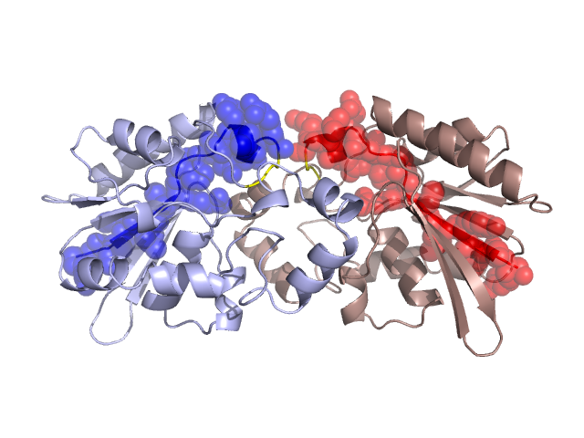
Pfam Domains mapped on to the structure: 1FBT No. Chain ID Pfam ID Pfam Description Linkout - Pfam Linkout - CDD 1 A PF00300 Histidine phosphatase superfamily (branch 1) PF00300 PF00300 Conserved Domain Database Superfamily Annotations: 1FBT No. PDB ID PSSM ID CDD Accession Superfamily Short Name Linkout - CDD 1 1FBT 132718 cd07067 HP_PGM_like - cl11399 2 1FBT 212312 cl11399 HP superfamily - - Structural Details of PDB entry 1FBT Structural Details of PDB entry 1FBT PDBid Chains Hinge Swapped Domain 1FBT A,B A:17-19,B:17-19 A:1-16,B:1-16 Swapped-domain interface residues and interactions: Chains Residues A 7, 14, 15, 16, 17, 89, B 7, 14, 15, 16, 17, 89, Non-swapped-domain interface residues and interactions: Chains Residues A 18, 28, 87, 88, 90, 92, 93, 94, 96, 97, 100, 103, 143, 163, 165, 167, 168, 187, 190, B 18, 28, 87, 88, 92, 93, 94, 96, 100, 103, 143, 147, 163, 165, 167, 168, 187, Swapped domains are represented using trasperent spheres. Non-swapped part is represented using light color and cartoon representation. Hinge region is shown in yellow color. Mutations in critical regions: Chains Hinge Domain swapped interface Non-swapped interface Swapped Domain ANo mutationNo mutationNo mutationNo mutation BNo mutationNo mutationNo mutationNo mutation HIDE output: Homologues found through HIDE algorithm JMOL Visualization: 2D-plot: A:1FBT B:1FBT JOY Structural annotation for hinge hinge and swapped domain: Hinge Swapped domain JOY output: ali file:1FBT.ali atm file:1FBT.atm cof file:1FBT.cof hbd file:1FBT.hbd html file:1FBT.html pdb file:1FBT.pdb ps file:1FBT.ps psa file:1FBT.psa rtf file:1FBT.rtf sst file:1FBT.sst tem file:1FBT.tem