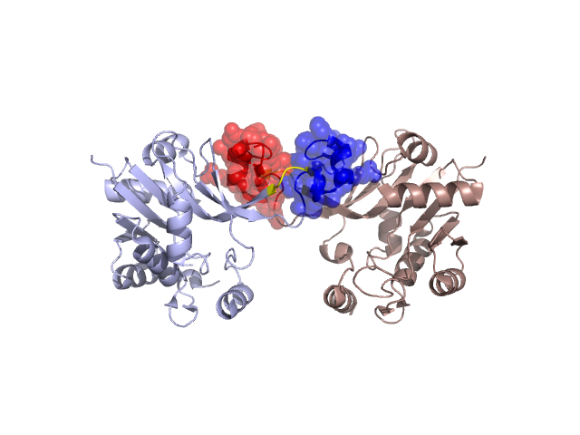
Structural Details of PDB entry 1EZI
Structural Details of PDB entry 1EZI
PDBid Chains Hinge Swapped Domain
1EZI
A,B
A:135-138,B:135-138;A:168-171,B:168-171
A:139-167,B:139-167
Swapped-domain interface residues and interactions:
Chains Residues
A
140 , 141 , 142 , 143 , 144 , 145 , 146 , 152 , 158 , 161 , 162 , 169 , 170 , 171 , 172 , 173 , 193 , 198 , 199 ,
B
140 , 141 , 142 , 143 , 144 , 145 , 146 , 150 , 152 , 158 , 161 , 162 , 169 , 170 , 171 , 172 , 173 , 174 , 193 , 197 ,
Non-swapped-domain interface residues and interactions:
Chains Residues
A
138 , 174 , 191 , 192 , 195 , 197 , 201 , 224 ,
B
138 , 191 , 192 , 195 , 196 , 199 , 201 ,