Pfam Domains mapped on to the structure: 1EW2
No.
Chain ID
Pfam ID
Pfam Description
Linkout - Pfam
Linkout - CDD
1
A
PF00245
Alkaline phosphatase
PF00245
PF00245
Conserved Domain Database Superfamily Annotations: 1EW2
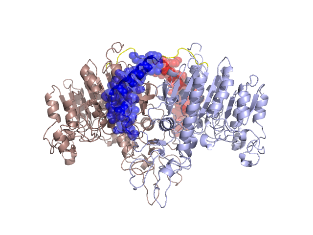
Structural Details of PDB entry 1EW2
Structural Details of PDB entry 1EW2
PDBid Chains Hinge Swapped Domain
1EW2
A,B
A:27-34,B:27-34
A:1-26,B:1-26
Swapped-domain interface residues and interactions:
Chains Residues
A
2 , 6 , 7 , 9 , 12 , 13 , 15 , 16 , 17 , 19 , 20 , 23 , 24 , 26 , 85 , 86 , 102 , 103 , 104 , 113 , 114 , 115 , 448 , 456 , 457 , 460 ,
B
2 , 6 , 7 , 9 , 12 , 13 , 15 , 16 , 17 , 19 , 20 , 23 , 24 , 26 , 85 , 86 , 102 , 103 , 104 , 113 , 114 , 115 , 448 , 456 , 457 , 460 ,
Non-swapped-domain interface residues and interactions:
Chains Residues
A
46 , 47 , 50 , 53 , 54 , 65 , 66 , 72 , 75 , 77 , 79 , 81 , 83 , 87 , 88 , 89 , 107 , 117 , 129 , 361 , 363 , 364 , 365 , 366 , 367 , 368 , 369 , 370 , 372 , 382 , 383 , 384 , 388 , 392 , 394 , 395 , 399 , 401 , 404 , 405 , 406 , 423 , 424 , 425 , 426 , 427 , 428 , 431 , 432 , 433 , 434 , 435 , 436 , 438 , 440 , 447 , 450 , 451 , 452 , 454 , 464 , 471 , 479 ,
B
46 , 47 , 50 , 53 , 54 , 65 , 66 , 72 , 75 , 77 , 79 , 81 , 83 , 87 , 88 , 89 , 107 , 117 , 129 , 361 , 363 , 364 , 365 , 366 , 367 , 368 , 369 , 370 , 372 , 382 , 383 , 384 , 388 , 392 , 394 , 395 , 399 , 401 , 404 , 405 , 406 , 423 , 424 , 425 , 426 , 427 , 428 , 431 , 432 , 433 , 434 , 435 , 436 , 438 , 440 , 447 , 450 , 451 , 452 , 454 , 464 , 471 ,
Mutations in critical regions:
Chains
Hinge
Domain swapped interface Non-swapped interface Swapped Domain
A No mutation No mutation No mutation No mutation B No mutation No mutation No mutation No mutation
HIDE output:
JMOL Visualization:
2D-plot:
JOY Structural annotation for hinge hinge and swapped domain:
JOY output: