Home BrowseSearch Help Group

Pfam Domains mapped on to the structure: 1ETK

No. Chain ID Pfam ID Pfam Description Linkout - Pfam Linkout - CDD
1 A PF02954 Bacterial regulatory protein, Fis family PF02954 PF02954

Conserved Domain Database Superfamily Annotations: 1ETK

No. PDB ID PSSM ID CDD Accession Superfamily Short Name Linkout - CDD
1 1ETK 212173 cl00088 HTH superfamily - -
Structural Details of PDB entry 1ETK

Structural Details of PDB entry 1ETK

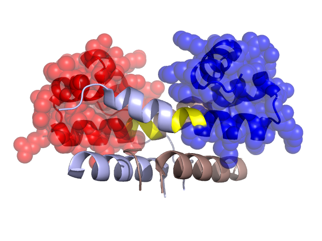
PDBid Chains Hinge Swapped Domain
1ETK A,B A:58-60,B:58-60 A:61-98,B:61-98

Swapped-domain interface residues and interactions:

Chains Residues
A 28, 31, 32, 35, 47, 48, 50, 51, 61, 62, 64, 65, 66, 69, 79, 80, 81, 82, 83, 91, 98,
B 28, 31, 32, 47, 48, 50, 51, 61, 62, 65, 66, 69, 79, 80, 81, 82, 83, 91,

Non-swapped-domain interface residues and interactions:

Chains Residues
A 10, 11, 12, 13, 27, 30, 33, 34, 36, 37, 38, 39, 41, 42, 53, 54, 55, 56, 57, 58, 59, 60,
B 5, 6, 7, 8, 10, 11, 12, 13, 27, 30, 33, 34, 35, 36, 37, 38, 39, 41, 53, 54, 55, 57, 58, 59,

Swapped domains are represented using trasperent spheres.
Non-swapped part is represented using light color and cartoon representation.
Hinge region is shown in yellow color.

Mutations in critical regions:

Chains Hinge Domain swapped interface Non-swapped interface Swapped Domain
ANo mutationNo mutationNo mutationALA(68)GLN,
BNo mutationNo mutationNo mutationALA(68)GLN,

HIDE output:

Homologues found through HIDE algorithm

JMOL Visualization:

Jmol_logo            Jmol_logo            Jmol_logo

2D-plot:

A:1ETK B:1ETK

JOY Structural annotation for hinge hinge and swapped domain:

Hinge Swapped domain

JOY output:

ali file:1ETK.ali
atm file:1ETK.atm
cof file:1ETK.cof
hbd file:1ETK.hbd
html file:1ETK.html
pdb file:1ETK.pdb
ps file:1ETK.ps
psa file:1ETK.psa
rtf file:1ETK.rtf
sst file:1ETK.sst
tem file:1ETK.tem