Home BrowseSearch Help Group

Pfam Domains mapped on to the structure: 1EP0

No. Chain ID Pfam ID Pfam Description Linkout - Pfam Linkout - CDD
1 A PF00908 dTDP-4-dehydrorhamnose 3,5-epimerase PF00908 PF00908

Conserved Domain Database Superfamily Annotations: 1EP0

No. PDB ID PSSM ID CDD Accession Superfamily Short Name Linkout - CDD
1 1EP0 212615 cl09118 Cupin_2 superfamily - -
Structural Details of PDB entry 1EP0

Structural Details of PDB entry 1EP0

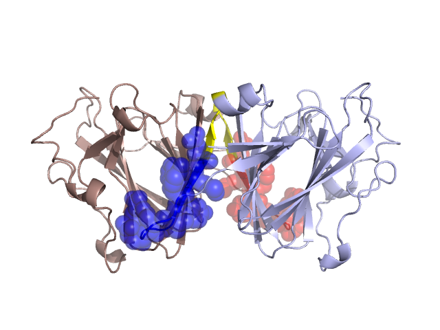
PDBid Chains Hinge Swapped Domain
1EP0 A,B A:19-21,B:19-21;A:32-36,B:32-36 A:22-31,B:22-31

Swapped-domain interface residues and interactions:

Chains Residues
A 25, 26, 27, 28, 29, 30, 31, 50, 51, 52, 53, 54, 56, 59, 167,
B 25, 26, 27, 28, 29, 30, 31, 50, 51, 52, 53, 54, 56, 59, 167,

Non-swapped-domain interface residues and interactions:

Chains Residues
A 32, 33, 34, 35, 36, 46, 47, 48, 49, 61, 76, 78, 79, 130, 132, 133, 134, 138, 169, 185,
B 32, 33, 34, 35, 36, 46, 47, 48, 49, 61, 76, 78, 79, 130, 132, 133, 134, 138, 169,

Swapped domains are represented using trasperent spheres.
Non-swapped part is represented using light color and cartoon representation.
Hinge region is shown in yellow color.

Mutations in critical regions:

Chains Hinge Domain swapped interface Non-swapped interface Swapped Domain
ANo mutationNo mutationNo mutationNo mutation
BNo mutationNo mutationNo mutationNo mutation

HIDE output:

Homologues found through HIDE algorithm

JMOL Visualization:

Jmol_logo            Jmol_logo            Jmol_logo

2D-plot:

A:1EP0 B:1EP0

JOY Structural annotation for hinge hinge and swapped domain:

Hinge Swapped domain

JOY output:

ali file:1EP0.ali
atm file:1EP0.atm
cof file:1EP0.cof
hbd file:1EP0.hbd
html file:1EP0.html
pdb file:1EP0.pdb
ps file:1EP0.ps
psa file:1EP0.psa
rtf file:1EP0.rtf
sst file:1EP0.sst
tem file:1EP0.tem