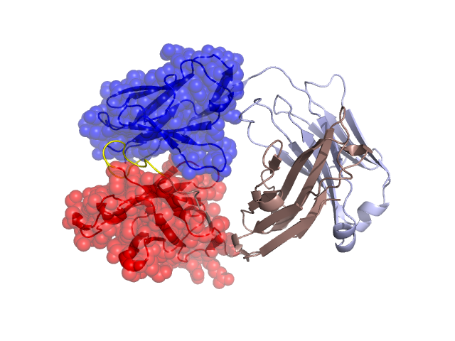
Structural Details of PDB entry 1CE1
Structural Details of PDB entry 1CE1
PDBid Chains Hinge Swapped Domain
1CE1
H,L
H:103-110,L:93-97
H:1-102,L:1-92
Swapped-domain interface residues and interactions:
Chains Residues
H
39 , 43 , 44 , 45 , 47 , 50 , 61 , 63 , 64 , 97 , 101 , 105 , 106 , 108 , 109 , 111 , 113 ,
L
1 , 34 , 36 , 38 , 41 , 43 , 44 , 46 , 49 , 55 , 87 , 89 , 91 , 95 , 96 , 98 , 100 ,
Non-swapped-domain interface residues and interactions:
Chains Residues
H
104 , 107 , 112 , 130 , 131 , 132 , 133 , 135 , 137 , 138 , 139 , 140 , 145 , 149 , 151 , 169 , 172 , 173 , 174 , 175 , 177 , 178 , 179 , 187 , 189 , 191 , 217 ,
L
94 , 114 , 116 , 117 , 118 , 119 , 121 , 123 , 124 , 127 , 129 , 131 , 133 , 135 , 137 , 138 , 160 , 161 , 162 , 163 , 164 , 165 , 167 , 174 , 176 , 178 , 180 , 207 , 208 , 209 , 211 ,