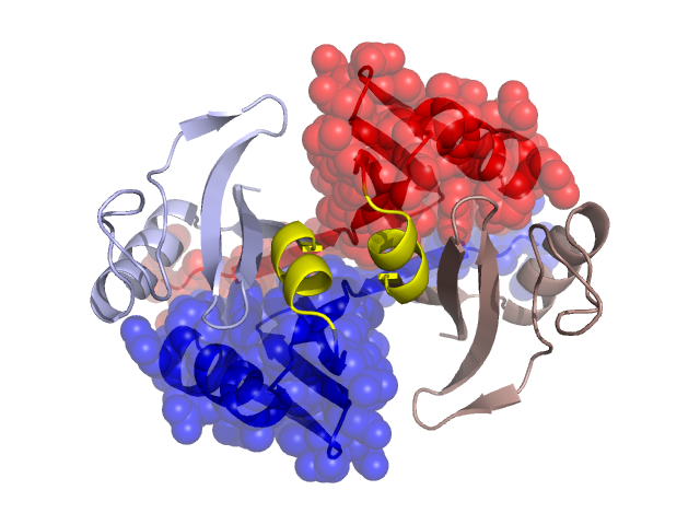
Structural Details of PDB entry 1BYL
Structural Details of PDB entry 1BYL
PDBid Chains Hinge Swapped Domain
1BYL
A,B
A:55-62,B:55-62
A:0-54,B:0-54
Swapped-domain interface residues and interactions:
Chains Residues
A
0 , 1 , 2 , 3 , 4 , 5 , 6 , 7 , 8 , 9 , 10 , 12 , 38 , 43 , 44 , 45 , 46 , 49 , 51 , 53 , 63 , 64 , 65 , 66 , 67 , 68 , 69 , 70 , 73 , 74 ,
B
0 , 1 , 2 , 3 , 4 , 5 , 6 , 7 , 8 , 9 , 10 , 12 , 38 , 43 , 44 , 45 , 46 , 49 , 51 , 53 , 63 , 64 , 65 , 66 , 67 , 68 , 69 , 70 , 73 , 74 ,
Non-swapped-domain interface residues and interactions:
Chains Residues
A
55 , 57 , 58 , 61 , 62 , 77 , 102 , 121 ,
B
55 , 57 , 58 , 61 , 62 , 77 , 102 ,