Home BrowseSearch Help Group

Pfam Domains mapped on to the structure: 1BUO

No. Chain ID Pfam ID Pfam Description Linkout - Pfam Linkout - CDD
1 A PF00651 BTB/POZ domain PF00651 PF00651

Gene Ontology Annotations: 1BUO

No. GO ID GO Description Linkout - AmiGO
1 GO:0005515 protein binding GO:0005515

Conserved Domain Database Superfamily Annotations: 1BUO

No. PDB ID PSSM ID CDD Accession Superfamily Short Name Linkout - CDD
1 1BUO 207624 cl02518 BTB superfamily - -
Structural Details of PDB entry 1BUO

Structural Details of PDB entry 1BUO

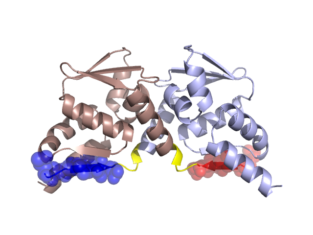
PDBid Chains Hinge Swapped Domain
1BUO A,B A:13-16,B:13-16 A:6-12,B:6-12

Swapped-domain interface residues and interactions:

Chains Residues
A 6, 7, 8, 9, 10, 11, 12, 90, 91, 92, 93, 94, 95, 96,
B 6, 7, 8, 9, 10, 11, 12, 90, 91, 92, 93, 94, 95, 96,

Non-swapped-domain interface residues and interactions:

Chains Residues
A 13, 16, 17, 18, 20, 21, 23, 24, 27, 32, 33, 35, 48, 49, 50, 51, 53, 54, 55, 60, 63, 86, 87, 99, 113, 117, 118, 121, 126,
B 13, 16, 17, 18, 20, 21, 23, 24, 27, 32, 33, 35, 48, 49, 50, 51, 53, 54, 55, 60, 63, 86, 87, 99, 113, 117, 118, 121,

Swapped domains are represented using trasperent spheres.
Non-swapped part is represented using light color and cartoon representation.
Hinge region is shown in yellow color.

Mutations in critical regions:

Chains Hinge Domain swapped interface Non-swapped interface Swapped Domain
ANo mutationNo mutationNo mutationNo mutation
BNo mutationNo mutationNo mutationNo mutation

HIDE output:

Homologues found through HIDE algorithm

JMOL Visualization:

Jmol_logo            Jmol_logo            Jmol_logo

2D-plot:

A:1BUO B:1BUO

JOY Structural annotation for hinge hinge and swapped domain:

Hinge Swapped domain

JOY output:

ali file:1BUO.ali
atm file:1BUO.atm
cof file:1BUO.cof
hbd file:1BUO.hbd
html file:1BUO.html
pdb file:1BUO.pdb
ps file:1BUO.ps
psa file:1BUO.psa
rtf file:1BUO.rtf
sst file:1BUO.sst
tem file:1BUO.tem