Pfam Domains mapped on to the structure: 1AQK
No.
Chain ID
Pfam ID
Pfam Description
Linkout - Pfam
Linkout - CDD
1
L
PF13927
Immunoglobulin domain
PF13927
PF13927
Conserved Domain Database Superfamily Annotations: 1AQK
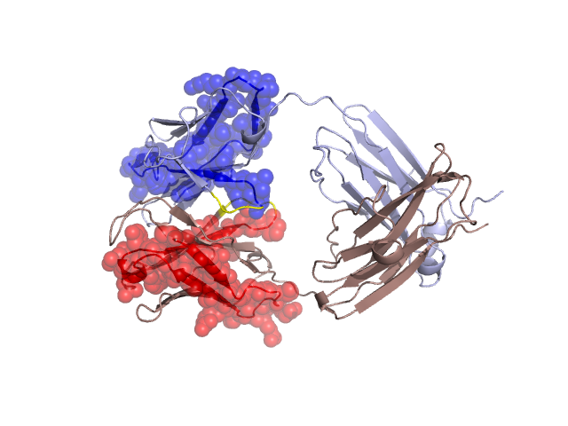
Structural Details of PDB entry 1AQK
Structural Details of PDB entry 1AQK
PDBid Chains Hinge Swapped Domain
1AQK
H,L
H:42-45,L:43-46
H:2-41,L:2-42
Swapped-domain interface residues and interactions:
Non-swapped-domain interface residues and interactions:
Chains Residues
H
44 , 45 , 47 , 57 , 59 , 95 , 100 , 101 , 106 , 107 , 108 , 110 , 111 , 113 , 114 , 115 , 132 , 133 , 134 , 135 , 139 , 140 , 147 , 151 , 153 , 174 , 176 , 177 , 179 , 181 , 182 , 187 , 189 , 191 , 219 ,
L
45 , 46 , 48 , 51 , 89 , 93 , 96 , 97 , 98 , 99 , 101 , 103 , 120 , 121 , 122 , 123 , 125 , 127 , 128 , 131 , 133 , 135 , 137 , 139 , 141 , 164 , 166 , 169 , 171 , 177 , 178 , 179 , 181 , 183 , 208 , 209 , 210 , 213 , 215 , 216 ,
Mutations in critical regions:
Chains
Hinge
Domain swapped interface Non-swapped interface Swapped Domain
H No mutation PRO(109)TYR, LYS(57)ASN, LEU(100)-, PHE(101)GLU, LEU(106)ARG, ALA(108)TYR, ASN(30)SER, ALA(33)GLY, ILE(34)MET, L No mutation PHE(33)HIS, THR(34)HIS, HIS(40)GLN, PHE(51)TYR, TYR(93)PHE, ALA(99)TRP, ALA(213)THR, ARG(17)LYS, VAL(18)ILE, THR(23)SER, SER(25)THR, ASN(26)SER, PHE(33)HIS, THR(34)HIS, HIS(40)GLN, LEU(41)VAL,
HIDE output:
JMOL Visualization:
2D-plot:
JOY Structural annotation for hinge hinge and swapped domain:
JOY output: