DSDBASE Help Central

DSDBASE Help Central |
|
Engineering Disuphide bonds |
|
| Search by PDB code | To search by PDB code, enter a valid PDB code (4 letter code starts with number), select the "PDB Code" option from radio button given), then submit the form.The result page contains PDB code (with link) and Protein name. User can reach the choosen entry by using link provided in PDB code. |
| Search by EC(Enzyme Commission) Number | To search by EC Number, enter a valid EC number (seperated
by period), select the "EC number" option from radio button
given), then submit the form.The result page contains PDB code (with link) ,EC number and Protein
name. User can reach the choosen entry by using link provided in PDB code. You may type 1 digit into the first field (the enzyme class), 1 or 2 digits into the second and third fields (subclass and sub-subclass), and up to 3 digits into the fourth field. For example, you may type 1.2.1.26 to get the corresponding ENZYME entry.You may also enter a partial EC number in order to get a list of all ENZYME entries whose EC numbers begin with the given pattern. For example, you may just type 1.2 |
| Search by Keyword | To search by keyword, enter keyword and select the "Key word" option from radio button given (by default this search engine will go for keyword search), then submit the form. The result page contains PDB code (with link) and Protein name. User can reach the choosen entry by using link provided in PDB code |
| Search by Sequence | To search by Sequence, Paste query sequence in FASTA format, then submit the form . The results contain PDB code (with link), alignment and corresponding scores (Blast output). User can reach the choosen entry by using link provided in PDB code. BLAST program used here is "blastall", database is "PDB-April2003" release. Standard E-value=10. In output PDB codes which has liks are present in DSDBASE, without links are absent. |
|| Back to top || || Blast page ||
MODIP online |
|
| Requirements for MODIP | There is a provision
to run MODIP online, for proteins not present in DSDBASE or for new protein structure 1. Upload or Paste the Coordinates file in PDB format fields. 2. PDB file can be single chain or multi chain. 3. MODIP won't accept 'C-alpha only' proteins. |
| Output | The
output is displayed on to the screen with information such as total number of disulphides,
number of modelled and native disulphides and grade distribution are given. followed by
this detailed results will be given in table. Table gives the information about Residues
in postion "i" and "j" which can accomadate disulphide bridge, and
corresponding grade.
|
|| Back to top || || MODIP online ||
Modelling Disulphide rich peptides |
Screen shot of DSDBASE search page
Search method |
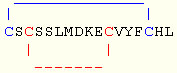
Ex : Endothelin ( PDB code : 1EDP ) Sequence : CSCSSLMDKECVYFCHL No of residues : 17 Disulphide bond connectivity : 1-15 , 3-11
|
Filling form for the above example
| 1. Select database : | Select database : |
Currently three different types of datasets are available.
| nr-db | a database of native and modelled disulphide bonds derived from non-redundant set of proteins (25% sequence identity, Hobohm et al.,1992) derived from PDB(April 2003). |
| nr-native | a database of only native disulphide bonds derived from non-redundant set of proteins (25% sequence identity, Hobohm et al.,1992) derived from PDB (April 2003). |
| fulldb(PDB-April 2003) | a database of native and modelled disulphide bonds derived from full PDB release(April 2003). |
| PDB-April 2001 | a database of native and modelled disulphide bonds derived from full PDB release(April 2001). |
Click here for statistics.
| 2. Protein name : | Protein Name : |
Enter any name (this is optional)
| 3. Sequence information : | |
This is optional. Enter the sequence of the protein/peptide. Please note that only the portion of protein/prptide whose connectivity is being searched is to be provided. In this example "CSCSSLMDKECVYFC" is to be pasted into the text area(the full length sequence is "CSCSSLMDKECVYFCHL"). Donot include charactres otherthan alphabets. No particular format is needed. Simply paste the raw sequence.
| 4. No of Disulphide bonds | No of Disulphide bonds : |
(user can chose from 1 - 5, based on the number of disulphide bonded system he/she wanted to search)
| 5. Enter disulphide bond connectivity of the query |
| Disulphide Bond 1 : Disulphide Bond 2 : Disulphide Bond 3 : Disulphide Bond 4 : Disulphide Bond 5 : |
|
( Enter only the residue numbers of pair involed in disulphide bond )
Since this example has only 2 disulphide bonds, leave the remaning boxes empty.
| 5. Advance options |
| a. Loop size relaxation : | Loop size relaxation : |
Explanation of loopsize relaxation
Now we can represent our connectivity pattern as follows
Loop 1: C..............C
1 15
In this case loop size = ( 15-1 ) + 1 = 15
If loop size is relaxed by 1 residue then search can be performed for loop size 15, 14
(15-1), 16 (15+1)
Loop 2: C........C
3 11
In this case loop size = ( 11-3 ) + 1 = 9
If loop size is relaxed by 1 residue then search can be performed for loop size 9, 8
(9-1), 10(9+1)
| b Loop proximity relaxation : | Loop proximity relaxation : |
Explanation of loop proximity relaxation
Loop proximity of given query can be represent like this
C.C.......C...C
1 3 11
15
.
...
+1
-3
Spatial distance between starting residue of bond1 and bond2
= 3 - 1 = +1
Spatial distance between end residue of bond1 and bond2
= 11 -15 = -3
By default (even without invoking this option), the search engine looks
for any sub-structural motif of a protein having loop sizes similar to query and also
their spatial distances.
If spacial distance is relaxed by 1 residue then,
spatial distance for starting residue : +1, 0
(1-1), 2 (1+1)
spatial distance for end residue : -3, -4
(-3-1), -2 (-3+1)
and search can be performed as follows,
| C.C.......C...C 1 3 11 15 . ... +1 -3 |
[ OR ] | C..C.......C..C 1 4 12 15 .. .. +2 -2 |
[ OR ] | CC.......C....C 12 10 15 .... 0 -4 |
|| Back to top || || Modelling page ||
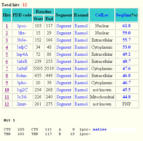
The output file contains following informations.
Screen shot of search result for Endothelin
A click on ......
Hit takes you to the substructure information (see below for more on this) PDB code leads to the corresponding entry in DSDBASE. Segment gives PDB file for Given region alone ( Example: for Hit 1 segment gives PDB file for residue no 15 to 29 ) CelLoc CelLoc is the subcellular location of the protein based on the aminiacid sequence alone. The results are from the SubLoc prediction server. Ref-Support vector machine approach for protein subcellular localization prediction.Bioinformatics. 2001 Aug;17(8):721-8. PMID: 11524373 [PubMed - indexed for MEDLINE]
"Not Known" - SubLoc couldn't predict the cellular location of the protein.SeqSim SeqSim is the the percentage of sequence similarity of the hit with the query sequence. Note that the cysteine positions are not fixed. INS = Input Not Supplied. FNP = File Not Present: Most probably this may be an obsolete/theoretical entry in the PDB. Rasmol option to veiw that particular region alone in Rasmol viewer A 'Hit' is ...............
CYS - 17 CYS - 25 C 9 1fre a b c d e f g h i
a amino acid in ith position b chain identifier c amino acid number of ith position d amino acid in jth position e chain identifier f amino acid number of jth position g grade h loopsize i PDB code
Native represents a substructural connectivity where both the residues are
CYS(Cysteine). In most instances these are as annotated in the PDB file and here
represented in Blue color. If MODIP only predicts these
connectivities, they are represented in Red color.
Redox-active represents redox-active disulphide bonds which are functionally
important and expected to behave differently from typical structural disulphide bonds.
These are reductive, reaction probably involves nucleophilic attack by the Cys thiolate on
the substrate to form a mixed disulphide intermediate.
|| Back to top || || Modelling page ||
Rasmol |
RasMol2 is a molecular graphics program intended for the visualisation of proteins, nucleic acids and small molecules. The program is aimed at display, teaching and generation of publication quality images. RasMol runs on Microsoft Windows, Apple Macintosh, UNIX and VMS systems. The UNIX and VMS systems require an 8, 24 or 32 bit colour X Windows display (X11R4 or later). The program reads in a molecule co-ordinate file and interactively displays the molecule on the screen in a variety of colour schemes and molecule representations. Currently available representations include depth-cued wireframes, 'Dreiding' sticks, spacefilling (CPK) spheres, ball and stick, solid and strand biomolecular ribbons, atom labels and dot surfaces.
Get a complete source code of rasmol and install it in your system.
Configure your browser as follows : Go to (In netscape) Edit --> Preferences --> Navigator --> Applications --> New
Fill the following : Description : PDB file; MIME Type : chemical/x-pdb;
Activate the option Application : Application :"rasmol_path" ("rasmol_path" = Rasmol path in your system.)
|| Back to top ||

 Rasmol option to view mutant pdb file also available.